Solved The X Ray Image Shown Here Shows Of The Principle Chegg

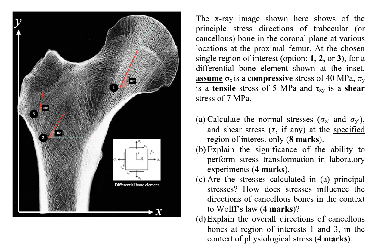
Solved The X Ray Image Shown Here Shows Of The Principle Chegg The x ray image shown here shows of the principle stress directions of trabecular (or cancellous) bone in the coronal plane at various locations at the proximal femur. at the chosen single region of interest (option: 1 , 2 , or 3 ), for a differential bone element shown at the inset, assume σ x is a compressive stress of 40 mpa , σ y is a tensile stress of 5 mpa and τ xy is a shear stress. Answer to solved the x ray image shown here shows of the principle | chegg.

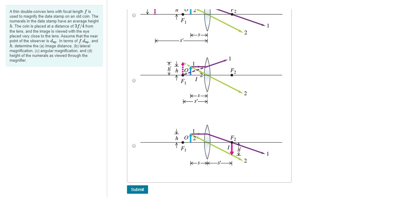
Solved Part A Select The Correct Principal Ray Diagram Chegg After wilhelm roentgen discovered x rays in 1895, william henry bragg pioneered the determination of crystal structure by x ray diffraction methods, began a lifelong investigation of the nature of radiation, principally x rays but also alpha and beta particles and gamma rays. after the discovery of the diffraction of x rays by crystals in 1912. The animation below shows ray tracing with two principle rays shown for the scenario of an object placed between the mirror and the focal point. since the object is to the right of the focal point and the center of curvature, the principle rays that would be going through those points to reach the mirror are now the rays that are coming from the direction of the these two points toward the mirror. Key points. x rays are produced within the x ray machine, also known as an x ray tube. no external radioactive material is involved. radiographers can change the current and voltage settings on the x ray machine in order to manipulate the properties of the x ray beam produced. different x ray beam spectra are applied to different body parts. The brachistochrone problem was one of the earliest problems posed in the calculus of variations. newton was challenged to solve the problem in 1696, and did so the very next day (boyer and merzbach 1991, p. 405). in fact, the solution, which is a segment of a cycloid, was found by leibniz, l'hospital, newton, and the two bernoullis.

Solved Draw A Principle Ray Diagram To Locate The Image Chegg Key points. x rays are produced within the x ray machine, also known as an x ray tube. no external radioactive material is involved. radiographers can change the current and voltage settings on the x ray machine in order to manipulate the properties of the x ray beam produced. different x ray beam spectra are applied to different body parts. The brachistochrone problem was one of the earliest problems posed in the calculus of variations. newton was challenged to solve the problem in 1696, and did so the very next day (boyer and merzbach 1991, p. 405). in fact, the solution, which is a segment of a cycloid, was found by leibniz, l'hospital, newton, and the two bernoullis. X rays. the study of atomic energy transitions enables us to understand x rays and x ray technology. like all electromagnetic radiation, x rays are made of photons. x ray photons are produced when electrons in the outermost shells of an atom drop to the inner shells. (hydrogen atoms do not emit x rays, because the electron energy levels are too. The ray tracing to scale in figure 9 shows two rays from a point on the bulb’s filament crossing about 1.50 m on the far side of the lens. thus the image distance \(d {i}\) is about 1.50 m. similarly, the image height based on ray tracing is greater than the object height by about a factor of 2, and the image is inverted. thus \(m\) is about.

Solved Consider The Ray Diagram Shown In The Figure Chegg X rays. the study of atomic energy transitions enables us to understand x rays and x ray technology. like all electromagnetic radiation, x rays are made of photons. x ray photons are produced when electrons in the outermost shells of an atom drop to the inner shells. (hydrogen atoms do not emit x rays, because the electron energy levels are too. The ray tracing to scale in figure 9 shows two rays from a point on the bulb’s filament crossing about 1.50 m on the far side of the lens. thus the image distance \(d {i}\) is about 1.50 m. similarly, the image height based on ray tracing is greater than the object height by about a factor of 2, and the image is inverted. thus \(m\) is about.

Solved Figure 1 Shows The Principal Ray Diagram Of The Chegg

Comments are closed.