Posterior Muscles Of The Shoulder Girdle And Shoulder Joint Diagram

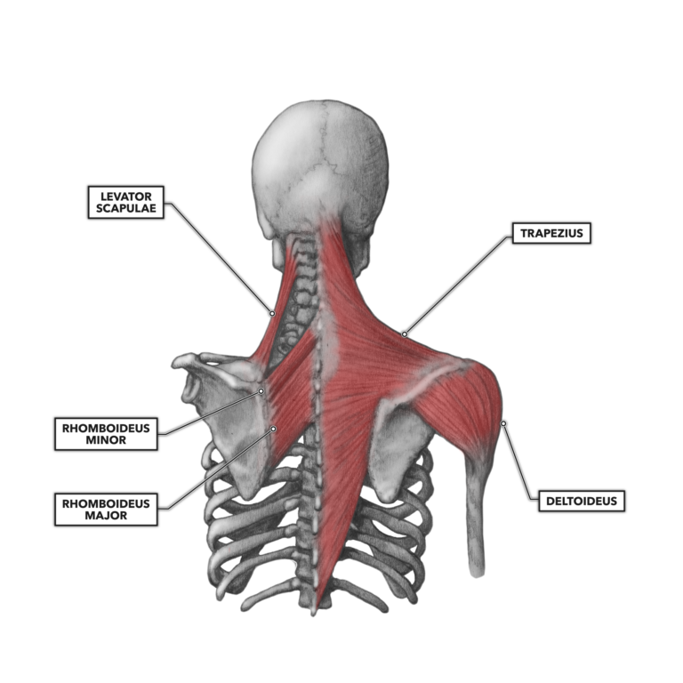
Muscles Of The Shoulder Posterior Modernheal The shoulder girdle functions as the anchor that attaches the upper limbs to the axial skeleton. additionally, the shoulder girdle allows for a large range of motion, mainly in the highly mobile scapulothoracic joint. this article will discuss the anatomy and functions of the shoulder girdle. key facts about the shoulder girdle. The muscles of the shoulder support and produce the movements of the shoulder girdle. they attach the appendicular skeleton of the upper limb to the axial skeleton of the trunk. four of them are found on the anterior aspect of the shoulder, whereas the rest are located on the shoulder’s posterior aspect and in the back.

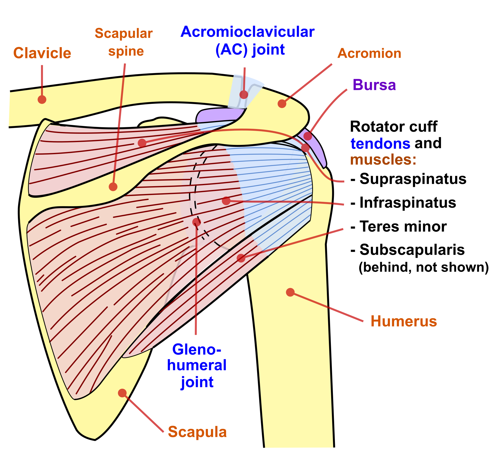
Shoulder Girdle вђ Anatomy Tutorial Medical Radiology Canada Winnipeg The shoulder joint is formed by an articulation between the head of the humerus and the glenoid cavity (or fossa) of the scapula. this gives rise to the alternate name for the shoulder joint – the glenohumeral joint. like most synovial joints, the articulating surfaces are covered with hyaline cartilage. the head of the humerus is much larger. The shoulder joint is protected superiorly by an arch, which is formed by the coracoid process of the scapula, the acromion process of the scapula and the clavicle. it is an extremely mobile joint, in which stability has been sacrificed for mobility. the bones of the pectoral girdle (clavicle and scapula) provide increased mobility to the. Bones, ligaments, muscles and movements of the shoulder joint. the glenohumeral, or shoulder, joint is a synovial joint that attaches the upper limb to the axial skeleton. it is a ball and socket joint, formed between the glenoid fossa of scapula (gleno ) and the head of humerus ( humeral). acting in conjunction with the pectoral girdle, the. The shoulder joint, also known as the glenohumeral joint, is a ball and socket joint with the most extensive range of motion in the human body. the shoulder muscles have a wide range of functions, including abduction, adduction, flexion, extension, internal and external rotation.[1] the central bony structure of the shoulder is the scapula, where all of the muscles interact. at the lateral.

Rotator Cuff Shoulder Muscle Anatomy At Timothy Guzman Blog Bones, ligaments, muscles and movements of the shoulder joint. the glenohumeral, or shoulder, joint is a synovial joint that attaches the upper limb to the axial skeleton. it is a ball and socket joint, formed between the glenoid fossa of scapula (gleno ) and the head of humerus ( humeral). acting in conjunction with the pectoral girdle, the. The shoulder joint, also known as the glenohumeral joint, is a ball and socket joint with the most extensive range of motion in the human body. the shoulder muscles have a wide range of functions, including abduction, adduction, flexion, extension, internal and external rotation.[1] the central bony structure of the shoulder is the scapula, where all of the muscles interact. at the lateral. Fig 7 • posterior muscles of the shoulder girdle. (1) rhomboids; (2) levator scapulae; (3) trapezius; (4) deltoid; (5) latissimus dorsi. the movements that occur when the shoulder is moved and that can be assessed in shoulder girdle examination are: elevation: the shoulder moves upwards in the frontal plane. The shoulder is structurally and functionally complex as it is one of the most freely moveable areas in the human body due to the articulation at the glenohumeral joint. it contains the shoulder girdle, which connects the upper limb to the axial skeleton via the sternoclavicular joint. the high range of motion of the shoulder comes at the expense of decreased stability of the joint, and it is.

Crossfit Shoulder Muscles Part 2 Posterior Musculature Fig 7 • posterior muscles of the shoulder girdle. (1) rhomboids; (2) levator scapulae; (3) trapezius; (4) deltoid; (5) latissimus dorsi. the movements that occur when the shoulder is moved and that can be assessed in shoulder girdle examination are: elevation: the shoulder moves upwards in the frontal plane. The shoulder is structurally and functionally complex as it is one of the most freely moveable areas in the human body due to the articulation at the glenohumeral joint. it contains the shoulder girdle, which connects the upper limb to the axial skeleton via the sternoclavicular joint. the high range of motion of the shoulder comes at the expense of decreased stability of the joint, and it is.

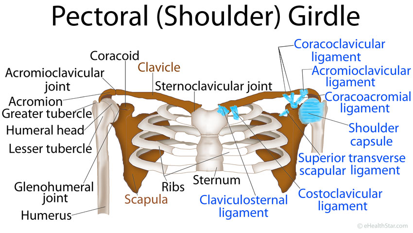
Pectoral Girdle Anatomy Bones Muscles Function Diagram Ehealthstar

Comments are closed.