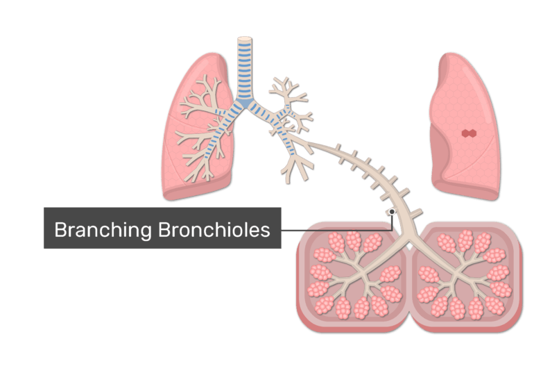
Nroer File Image Structure Of Affected Bronchioles

Nroer File Image Structure Of Affected Bronchioles The bronchioles are air passages in the lungs that deliver air to the alveoli. they can be affected by a number of different conditions, from asthma to cystic fibrosis. infection with bacteria or viruses can also lead to conditions like bronchopneumonia. conditions of the bronchioles are treated according to their cause. The bronchioles are part of conducting zone of the respiratory system. the conducting zone allows air to travel from the trachea into the alveoli, where gaseous exchange occurs. the bronchioles start off as bronchi. the right and left main bronchi branch off from the trachea into the lungs. subsequently, they further branch into smaller bronchi.

Structure Of Bronchioles Bronchi. bronchi are plural for bronchus and represent the passageways leading into the lungs. the first bronchi branch from trachea, and they are the right and left main bronchi. these bronchi are the widest and they enter the lung. after entering the lungs, the bronchi continue to branch further into the secondary bronchi, known as lobar. The trachea, bronchi and bronchioles form the tracheobronchial tree – a system of airways that allow passage of air into the lungs, where gas exchange occurs. these airways are located in the neck and thorax. in this article we will look at the anatomical position, structure and neurovascular supply of the airways; as well as considering. Respiratory bronchioles. terminal bronchioles then branch to form respiratory bronchioles. this marks the transition point from the conducting to the respiratory portion of the respiratory system. the epithelium here is simple cuboidal that may be ciliated proximally, but devoid of cilia distally. the smooth muscle layer is thinner here than in. The organs of the respiratory system form a continuous system of passages called the respiratory tract, through which air flows into and out of the body. the respiratory tract has two major divisions: the upper respiratory tract and the lower respiratory tract. the organs in each division are shown in figure 16.2.2 16.2.
Bronchi What Are They Location Structure Anatomy Func Vrogue Co Respiratory bronchioles. terminal bronchioles then branch to form respiratory bronchioles. this marks the transition point from the conducting to the respiratory portion of the respiratory system. the epithelium here is simple cuboidal that may be ciliated proximally, but devoid of cilia distally. the smooth muscle layer is thinner here than in. The organs of the respiratory system form a continuous system of passages called the respiratory tract, through which air flows into and out of the body. the respiratory tract has two major divisions: the upper respiratory tract and the lower respiratory tract. the organs in each division are shown in figure 16.2.2 16.2. Figure 19.5.1 19.5. 1: trachea (a) the tracheal tube is formed by stacked, c shaped pieces of hyaline cartilage. (b) the layer visible in this cross section of tracheal wall tissue between the hyaline cartilage and the lumen of the trachea is the mucosa, which is composed of pseudostratified ciliated columnar epithelium that contains goblet cells. Summary. the bronchioles are the smallest air passages in the lungs and they end in tiny sacs called alveoli. the wall of each bronchiole has a layer of smooth muscle that can contract to narrow the airway. distally are the smallest bronchioles called terminal bronchioles, which give rise to the respiratory bronchioles that end up with alveolar.

Bronchioles Definition Location Anatomy Function Diag Vrogue Co Figure 19.5.1 19.5. 1: trachea (a) the tracheal tube is formed by stacked, c shaped pieces of hyaline cartilage. (b) the layer visible in this cross section of tracheal wall tissue between the hyaline cartilage and the lumen of the trachea is the mucosa, which is composed of pseudostratified ciliated columnar epithelium that contains goblet cells. Summary. the bronchioles are the smallest air passages in the lungs and they end in tiny sacs called alveoli. the wall of each bronchiole has a layer of smooth muscle that can contract to narrow the airway. distally are the smallest bronchioles called terminal bronchioles, which give rise to the respiratory bronchioles that end up with alveolar.

Respiratory System Bronchioles Vrogue Co

Comments are closed.