Mri Imaging Of Cervical Spine Shorts

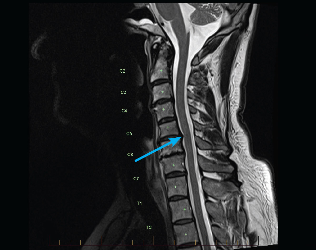
Normal Cervical Spine Mri Image Radiopaedia Org When it comes to understanding cervical spine health, magnetic resonance imaging (mri) plays an important role in diagnosing and distinguishing between normal and abnormal conditions. the cervical spine consists of seven vertebrae (c1 c7) located in the neck region and houses the spinal cord, supporting the head’s weight and allowing for. Megan soliman, md. cervical spine mri (magnetic resonance imaging) is a useful tool when people have symptoms such as neck pain or numbness in their arms. it can detect a variety of medical conditions including bulging discs, herniated discs, tumors, aneurysms, and some autoimmune disorders. unlike an x ray or ct scan, it poses no radiation risk.

Adult Mri Series Mri Of Cervical Spine Radiculopathy An mri of the cervical spine is usually conducted with the patient in the supine position. technical parameters coil. head and neck coil. scan geometry. in plane spatial resolution: ≤0.7 x 0.7 mm. field of view (fov): 200 240 (sagittal coronal) 100 160 (axial) slice thickness: ≤3mm 2 4. planning. a typical mri of the cervical spine might. The average cervical spinal canal sagittal diameter on mri is approximately 13.7 14.1 mm [34,35]. a cervical spinal canal sagittal diameter of < 13 mm is considered relatively narrowed, and < 10 mm as absolutely narrowed, based on measurements obtained from cervical spine radiographs with correlation of myelopathy symptoms . mri has the. A cervical spine mri scan is used to help diagnose: tumors in your bones or soft tissues. bulging discs, or herniated discs. aneurysms, which are bulges in arteries, or other vascular disorders. Magnetic resonance imaging (mri) is instrumental in evaluation of symptomatic cervical spine degeneration and guidance of surgical treatment. in the setting of trauma, mri can help define unexplained neurologic injuries, assess for soft tissue damage, and exclude cervical spine injury. it has also become extremely beneficial in the diagnosis.

Normal Cervical Spine Mri Explained Dr Jeffrey P Johnson Hd Youtube A cervical spine mri scan is used to help diagnose: tumors in your bones or soft tissues. bulging discs, or herniated discs. aneurysms, which are bulges in arteries, or other vascular disorders. Magnetic resonance imaging (mri) is instrumental in evaluation of symptomatic cervical spine degeneration and guidance of surgical treatment. in the setting of trauma, mri can help define unexplained neurologic injuries, assess for soft tissue damage, and exclude cervical spine injury. it has also become extremely beneficial in the diagnosis. The normal cervical spine canal should be between 10 14 millimeters. some report even larger, up to 16 millimeters. sometimes this measurement is included in a report. the spinal cord itself extends through the entire cervical spine. at each disc level, a nerve exits the spine and goes to a specific region of the arm. Diagnosis of cervical spine injury in patients following trauma involves imaging. cervical spine injuries can range from those that are minor and stable to more severe injuries that involve vertebral fractures or damage to the spinal cord, nerve root, ligaments, or vessels. this topic describes cervical spine imaging in adults including the.

Normal Trauma Cervical Spine Mri Radiology Case Radiopaedia Org The normal cervical spine canal should be between 10 14 millimeters. some report even larger, up to 16 millimeters. sometimes this measurement is included in a report. the spinal cord itself extends through the entire cervical spine. at each disc level, a nerve exits the spine and goes to a specific region of the arm. Diagnosis of cervical spine injury in patients following trauma involves imaging. cervical spine injuries can range from those that are minor and stable to more severe injuries that involve vertebral fractures or damage to the spinal cord, nerve root, ligaments, or vessels. this topic describes cervical spine imaging in adults including the.

Comments are closed.