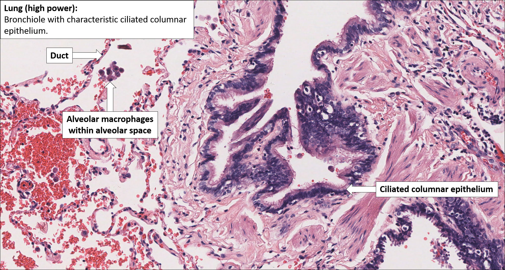
Lung Histology Diagram

Respiratory System The lungs are a pair of primary organs of respiration, present in the thoracic cavity beside the mediastinum. they are covered by a thin double layered serous membrane called the pleura. the respiratory system consists of two components, the conducting portion, and the respiratory portion. the conducting portion brings the air from outside to. Type ii dome shaped, cuboidal epithelial cells that project into the lumen. secrete surfactant that covers the alveolar surface and reduces surface tension. macrophages (or dust cells) (. #1. , #2. and. #3. ) large, dark cells within alveoli that engulf dust particles, bacteria, and other pathogens.

Lung Histology Diagram The respiratory system consists of several organs working together to facilitate the exchange of oxygen and carbon dioxide between the air and the bloodstream, supporting cellular respiration. the respiratory tract is divided into two sections; the upper and l ower respiratory tract. the upper respiratory tract includes the paranasal sinuses. Anatomical position and relations. the lungs lie either side of the mediastinum, within the thoracic cavity. each lung is surrounded by a pleural cavity, which is formed by the visceral and parietal pleura. they are suspended from the mediastinum by the lung root – a collection of structures entering and leaving the lungs. Anatomy and histology of the lung joseph f. tomashefski, jr., and carol f. farver the lung is uniquely designed to accomplish its major functions of movement of air and the delivery of oxygen to and removal of carbon dioxide from the circul ation. pulmonary anatomic compartments are tightly. Respiratory system. palate, h&e, 20x (respiratory epithelium lining nasal cavity, stratified squamous lining oral cavity). fetal face, frontal section, h&e, 40x (nasal cavity, nasal mucosa, respiratory epithelium, olfactory epithelium).

Lung Histology Diagram Anatomy and histology of the lung joseph f. tomashefski, jr., and carol f. farver the lung is uniquely designed to accomplish its major functions of movement of air and the delivery of oxygen to and removal of carbon dioxide from the circul ation. pulmonary anatomic compartments are tightly. Respiratory system. palate, h&e, 20x (respiratory epithelium lining nasal cavity, stratified squamous lining oral cavity). fetal face, frontal section, h&e, 40x (nasal cavity, nasal mucosa, respiratory epithelium, olfactory epithelium). The gaps between the rings of cartilage are filled by the trachealis muscle a bundle of smooth muscle, and fibroelastic tissue. together these hold the lumen of the trachea open, but allow flexibility during inspiration and expiration. the respiratory mucosa and submucosa are adapted to warm and moisten the air, and to trap particles in mucous. Introduction. knowledge of normal lung anatomy and function is important for the interpretation of lung biopsies and resections. an understanding of different cell structures and functions allows for a greater appreciation of disease states.

Comments are closed.