Lateral Malleolus Fibula Ankle Palpation

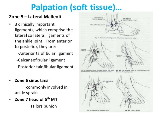
Lateral Malleolus Fibula Ankle Palpation First, have the patient lying down supine with the knee bent on the affected side. then, observe the lateral aspect of the foot and ankle for hematomas or bruises. then, locate the three lateral ligaments and palpate along their course for crepitus and tenderness. lateral ankle inspection and palpation. image credit. Laterally, palpation includes the tip of the lateral malleolus, the fibula, and the three lateral ligaments: anterior talofibular, posterior talofibular, and fibulocalcaneal. because an inversion injury of the ankle can fracture the proximal fibula, the proximal fibula is palpated. the base of the 5th metatarsal is also palpated.

Examination Of Foot And Ankle Percutaneous placement of syndesmosis screws. access to the posterolateral tibia. approach. position. supine with bump under buttock. incision. make longitudinal incision along the posterior margin of the fibula (center incision over fracture site) extend 2 cm distal to the tip of the lateral malleolus (if needed) superficial dissection. An impact during a car crash can also cause an ankle fracture. lateral malleolus fractures are characterized by the following signs and symptoms: immediate severe pain. swelling of the ankle. bruising. tenderness to the touch. inability to bear weight on the injured ankle. ankle deformity. Palpation along the entire length of the fibular is important in order to pick up signs of this injury as it can be seen in patients with relatively unremarkable ankle x rays; if the patient is discovered to have either a medial malleolus fracture or high fibular fracture on x ray, tibia fibula films are indicated to rule out this type of fracture. Palpation of the ankle should include the medial and lateral gutters, the tibiotalar plafond, the talar dome, and the syndesmosis. proximally at the ankle, the medial malleolus of the tibia and the lateral malleolus of the fibula are prominent and should always be palpable.

Lateral Malleolus Fibula Ankle Palpation Palpation along the entire length of the fibular is important in order to pick up signs of this injury as it can be seen in patients with relatively unremarkable ankle x rays; if the patient is discovered to have either a medial malleolus fracture or high fibular fracture on x ray, tibia fibula films are indicated to rule out this type of fracture. Palpation of the ankle should include the medial and lateral gutters, the tibiotalar plafond, the talar dome, and the syndesmosis. proximally at the ankle, the medial malleolus of the tibia and the lateral malleolus of the fibula are prominent and should always be palpable. • palpation – pain on palpation of the posterior aspect of the medial and lateral malleolus (both sites are void of ligamentous attachments), the base of the fifth metatarsal and the proximal fibula are suggestive of fracture (figure 3).2 lack of tenderness on palpation of the atfl excludes atfl rupture. Epidemiology. very common: ∼187 ankle fractures per 100,000 people each year (1) fractures to ankle or midfoot occur in <15% of ankle sprains. most ankle fractures are malleolar fractures: 60–70% are unimalleolar (lateral being most common), 15–20% are bimalleolar, and 7–12% are trimalleolar (medial, lateral, and posterior malleoli) (2).

Kin191 A Ch 5 Ankle Lower Leg Evaluation Fall 2007 • palpation – pain on palpation of the posterior aspect of the medial and lateral malleolus (both sites are void of ligamentous attachments), the base of the fifth metatarsal and the proximal fibula are suggestive of fracture (figure 3).2 lack of tenderness on palpation of the atfl excludes atfl rupture. Epidemiology. very common: ∼187 ankle fractures per 100,000 people each year (1) fractures to ankle or midfoot occur in <15% of ankle sprains. most ankle fractures are malleolar fractures: 60–70% are unimalleolar (lateral being most common), 15–20% are bimalleolar, and 7–12% are trimalleolar (medial, lateral, and posterior malleoli) (2).

Comments are closed.