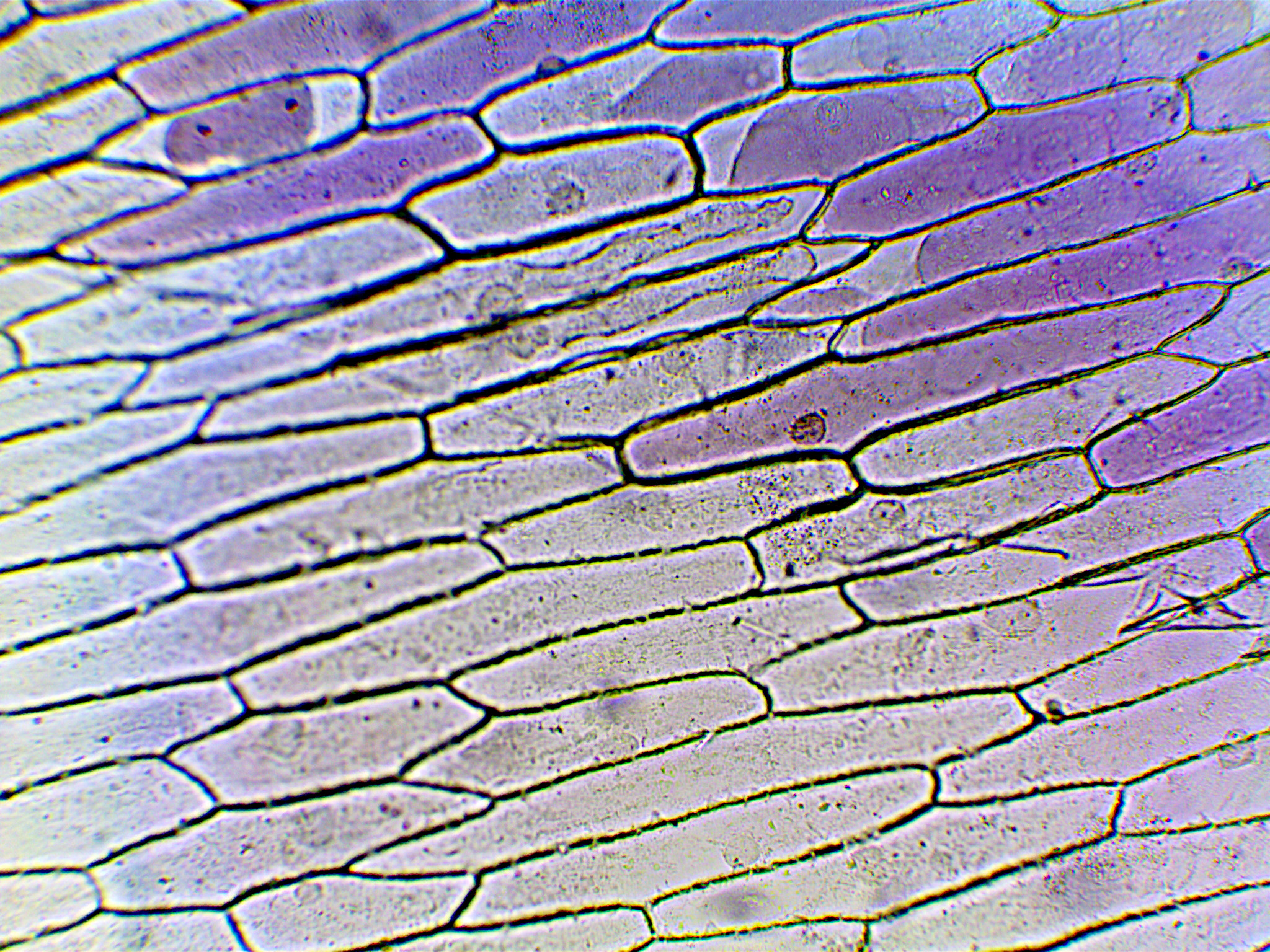
Labelled Diagram Of Onion Cell

Diagram Labeled Onion Cell Diagram Mydiagram Online Cell walls give structure. cell walls in plants are rigid, compared to other organisms. the cellulose present in the cell walls forms clearly defined tiles. in onion cells the tiles look very similar to rectangular bricks laid in offset runs. the rigid walls combined with water pressure within a cell provide strength and rigidity, giving plants. Observing onion cells under a microscope is a fun and easy activity for students and hobbyists alike. onion epidermal cells appear as a single thin layer and look highly organized and structured in terms of shape and size. certain parts of the cell are also clearly distinguishable with or without staining, making the activity even easier and.

Diagram Labeled Diagram Of An Onion Cell Mydiagram Online The onion peel cell experiment is a popular and educational activity used to observe and understand the structure of plant cells. this experiment focuses on the onion, a eukaryotic plant known for its multicellular composition. as we delve into this experiment, we explore the essential components that make up a cell, the building blocks of life. Add a drop of water at the center of the microscopic slide. having pulled of a thin membrane from the onion layer, lay it at the center of the microscopic slide (the drop of water will help flatten the membrane) add a drop of iodine solution on the onion membrane (or methylene blue) gently lay a microscopic cover slip on the membrane and press. The onion peel cell experiment is very popular for observing a plant cell structure. onion is a eukaryotic plant that contains multicellular cells. we know that the cell is a structural and functional unit of life that builds up living structures. the bulb of an onion is formed from modified leaves. like plant cells, onion cells have a rigid. Onion epidermal cell. these large cells from the epidermis of a red onion are naturally pigmented. the epidermal cells of onions provide a protective layer against viruses and fungi that may harm the sensitive tissues. because of their simple structure and transparency they are often used to introduce students to plant anatomy [1] or to.

Labelled Diagram Of Onion Cell The onion peel cell experiment is very popular for observing a plant cell structure. onion is a eukaryotic plant that contains multicellular cells. we know that the cell is a structural and functional unit of life that builds up living structures. the bulb of an onion is formed from modified leaves. like plant cells, onion cells have a rigid. Onion epidermal cell. these large cells from the epidermis of a red onion are naturally pigmented. the epidermal cells of onions provide a protective layer against viruses and fungi that may harm the sensitive tissues. because of their simple structure and transparency they are often used to introduce students to plant anatomy [1] or to. What would the onion cell(s) have looked like without the stain? 5. make a chart that lists the cell parts that you observed & labeled and tell what each part does for the cell. 6. describe where the cell parts were placed located inside the cell in relation to each other. 7. describe the ways that all the cells you observed were alike and how. Figure 10.3.1.1 10.3.1. 1: cells in an onion root in interphase and prophase. cell a has a large, dark nucleolus surrounded by greyish material (chromatin) that is enclosed within the nuclear membrane. a cell wall makes a box around each cell and the plasma membrane would be located just inside this box, though we cannot easily see it.

Diagram Labeled Onion Cell Diagram Mydiagram Online What would the onion cell(s) have looked like without the stain? 5. make a chart that lists the cell parts that you observed & labeled and tell what each part does for the cell. 6. describe where the cell parts were placed located inside the cell in relation to each other. 7. describe the ways that all the cells you observed were alike and how. Figure 10.3.1.1 10.3.1. 1: cells in an onion root in interphase and prophase. cell a has a large, dark nucleolus surrounded by greyish material (chromatin) that is enclosed within the nuclear membrane. a cell wall makes a box around each cell and the plasma membrane would be located just inside this box, though we cannot easily see it.
Labelled Diagram Of Onion Cell

Comments are closed.