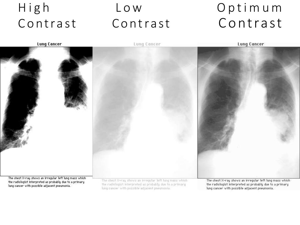
Ideal Radiography

Ideal Radiography Ppt The term “ideal image” throughout this website may refer to a corporate owned practice, franchisee, or physician owned practice. ideal image is the nation's leading medspa, partnering every client with a team of skin, face and body specialists and medical experts. we help people look and feel their best with non invasive treatments and. Ideal x ray beam characteristics the ideal x ray beam used for imaging should be: • sufficiently penetrating, to pass through the patient and react with the film emulsion or digital sensor and produce good contrast between the different shadows • parallel, i.e. non diverging, to prevent magnification of the image • produced from a point.

Ideal Radiographic Projection Ideal Radiographic Projection Ideal The term “ideal image” throughout this website may refer to a corporate owned practice, franchisee, or physician owned practice. ideal image is the nation's leading medspa, partnering every client with a team of skin, face and body specialists and medical experts. we help people look and feel their best with non invasive treatments and. In british english the word “ideal” means: “perfect, or “ best possible”. 1. john cameron in 1970 stated that, “there was negligible risk from x rays but many radiographs had poor image quality so that the risk from a false negative was significant”. image quality and patient exposure are 2 important aspects of radiography. The total effective dose of a chest x ray (in pa and lateral views) ranges from 0.06 to 0.25 msv, depending on the voltage of the system used and type of system (film screen or digital radiography). meanwhile, pa view accounts for 25% of the total effective dose of a chest x ray while lateral view accounts for 75% of the total effective dose 6 . Image quality can be defined as the attribute of the image that influences the clinician's certainty to perceive the appropriate diagnostic features from the image visually.[1][2] quality assurance or improvement is the proactive action to enhance the quality of care and services and cost effectively remove waste. this topic discusses the fundamental concepts of digital radiographic image.

Ideal Radiography The total effective dose of a chest x ray (in pa and lateral views) ranges from 0.06 to 0.25 msv, depending on the voltage of the system used and type of system (film screen or digital radiography). meanwhile, pa view accounts for 25% of the total effective dose of a chest x ray while lateral view accounts for 75% of the total effective dose 6 . Image quality can be defined as the attribute of the image that influences the clinician's certainty to perceive the appropriate diagnostic features from the image visually.[1][2] quality assurance or improvement is the proactive action to enhance the quality of care and services and cost effectively remove waste. this topic discusses the fundamental concepts of digital radiographic image. The ideal imaging system should permit a high quality image with minimal radiation exposure. dr has the potential to achieve this and further advances will possibly lead to lowering the radiation dose and using higher sensitivity plates to give good resolution and sharpness of images. portable radiography is another significant reason to adopt dr. Plain radiograph. a correctly placed nasogastric tube should 10: descend in the midline, following the path of the esophagus and avoiding the contours of the bronchi. clearly bisect the carina or bronchi. cross the diaphragm in the midline. have its tip visible below the left hemidiaphragm. ideally, the tip should be at least 10 cm beyond the.

Ideal Radiography Ppt The ideal imaging system should permit a high quality image with minimal radiation exposure. dr has the potential to achieve this and further advances will possibly lead to lowering the radiation dose and using higher sensitivity plates to give good resolution and sharpness of images. portable radiography is another significant reason to adopt dr. Plain radiograph. a correctly placed nasogastric tube should 10: descend in the midline, following the path of the esophagus and avoiding the contours of the bronchi. clearly bisect the carina or bronchi. cross the diaphragm in the midline. have its tip visible below the left hemidiaphragm. ideally, the tip should be at least 10 cm beyond the.

Ideal Radiography Ppt

Comments are closed.