Human Lung Organoid Label Free 3d Live Cell Imaging

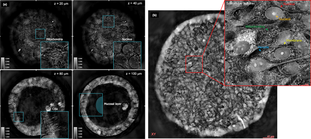
Human Lung Organoid Label Free 3d Live Cell Imaging Youtube In this paper, we demonstrate a label free high content imaging approach for observing changes in organoid morphology and structural changes occurring at the cellular and subcellular level. We show that an organoid with an initial cell number of eight increases at a low rate (average: 0.65 cells per hour) and reaches a maximum number of 107 cells after 6 days, whereas an organoid starting with nine cells increases at a high rate (average: 7.41 cells per hour) and ends up with 1077 cells after 6 days (fig. 3a, blue and green frames).

Holotomography Label Free 3d Imaging Of Live Cells And Organoids Ht is a 3d quantitative phase imaging technology [1] that can accurately measure the 3d refractive index (ri) distribution of unlabeled live cells. this has the same physical principles as x ray computed tomography (ct), although ct uses x rays to measure the 3d x ray absorbance distribution of the human body from multiple 2d images from. Non‐invasive label‐free imaging analysis pipeline for in situ characterization of 3d brain organoids. brain organoids provide a unique opportunity to model organ development in a system. Current imaging approaches limit the ability to perform multi scale characterization of three dimensional (3d) organotypic cultures (organoids) in large numbers. here, we present an automated. In this paper, we demonstrate a label free high content imaging approach for observing changes in organoid morphology and structural changes occurring at the cellular and subcellular level. enabled by microfluidic based culture of 3d cell systems and a novel 3d quantitative phase imaging method, we demonstrate the ability to perform non.

Holotomography Label Free 3d Imaging Of Live Cells And Organoids Current imaging approaches limit the ability to perform multi scale characterization of three dimensional (3d) organotypic cultures (organoids) in large numbers. here, we present an automated. In this paper, we demonstrate a label free high content imaging approach for observing changes in organoid morphology and structural changes occurring at the cellular and subcellular level. enabled by microfluidic based culture of 3d cell systems and a novel 3d quantitative phase imaging method, we demonstrate the ability to perform non. The goal of this current study is to reduce overall burden on assay duration and development in non small cell lung cancer (nsclc) organoids by leveraging label free multiphoton imaging. in this study, simultaneous label free autofluorescence multiharmonic (slam) microscopy was used to provide rich intracellular information based on endogenous. Correlative label free and fluorescence imaging systems have recently been reported (4, 11–14) for 3d imaging of organelles, cells, and tissues. building on these advancements, we have developed an automated microscope, named “mantis,” that synergizes light sheet and label free microscopy in multiwell plates.
3d Human Lung Alveolar Organoi Image Eurekalert Science News Releases The goal of this current study is to reduce overall burden on assay duration and development in non small cell lung cancer (nsclc) organoids by leveraging label free multiphoton imaging. in this study, simultaneous label free autofluorescence multiharmonic (slam) microscopy was used to provide rich intracellular information based on endogenous. Correlative label free and fluorescence imaging systems have recently been reported (4, 11–14) for 3d imaging of organelles, cells, and tissues. building on these advancements, we have developed an automated microscope, named “mantis,” that synergizes light sheet and label free microscopy in multiwell plates.

Deep Learning Based Image Analysis For Label Free Live Monitoring Of

Comments are closed.