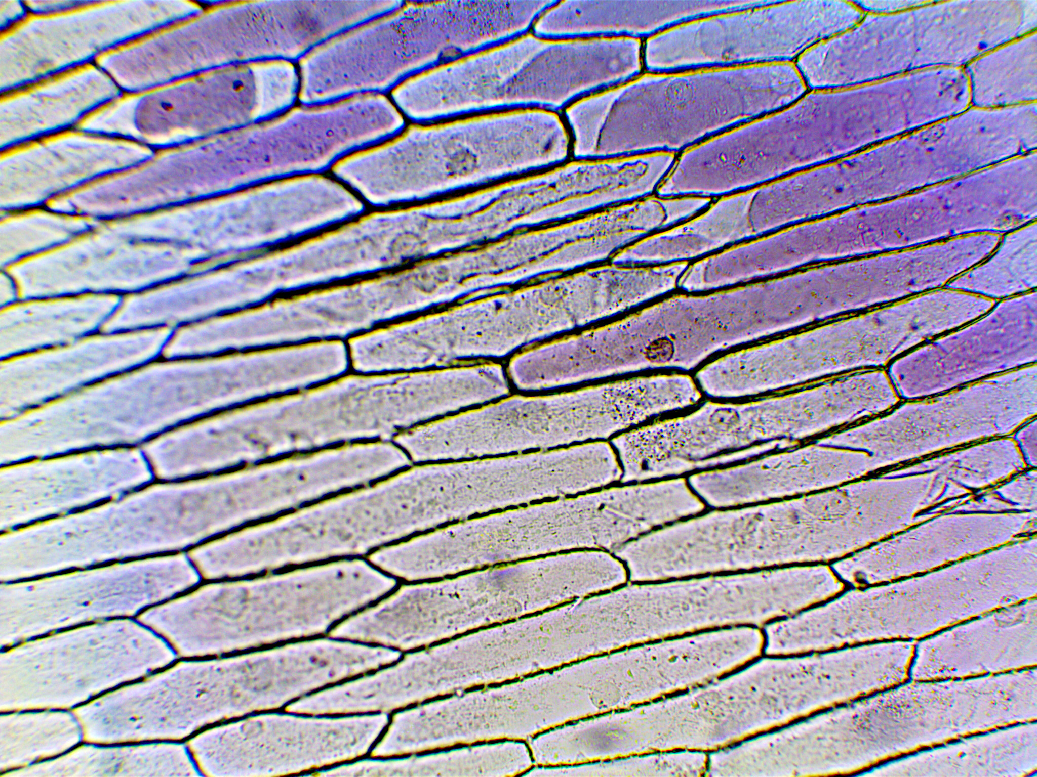
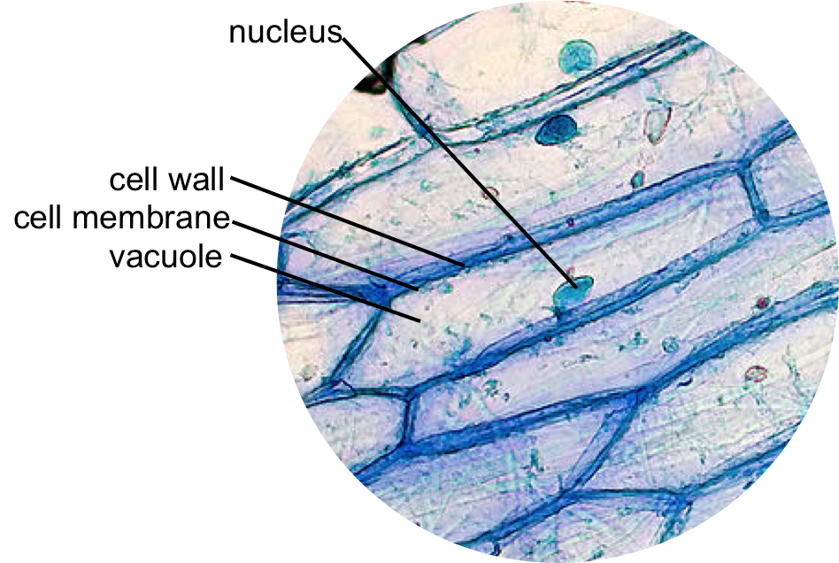
Diagram Of An Onion Cell Under A Microscope

Diagram Labeled Onion Cell Diagram Mydiagram Online Observing onion cells under a microscope is a fun and easy activity for students and hobbyists alike. onion epidermal cells appear as a single thin layer and look highly organized and structured in terms of shape and size. certain parts of the cell are also clearly distinguishable with or without staining, making the activity even easier and. Add a drop of water at the center of the microscopic slide. having pulled of a thin membrane from the onion layer, lay it at the center of the microscopic slide (the drop of water will help flatten the membrane) add a drop of iodine solution on the onion membrane (or methylene blue) gently lay a microscopic cover slip on the membrane and press.

Onion Cells Under Microscope The onion peel cell experiment is a popular and educational activity used to observe and understand the structure of plant cells. this experiment focuses on the onion, a eukaryotic plant known for its multicellular composition. as we delve into this experiment, we explore the essential components that make up a cell, the building blocks of life. Compound microscope; theory. an onion is a multicellular plant. the presence of a rigid cell wall and a large vacuole is a characteristic feature of a plant cell. thus, onion being a plant, comprises features common to plant cells. like plant cells, onion cells consist of a cell wall and cell membrane surrounding the cytoplasm, nucleus and a. One of the easiest labs in cell biology is observing onion cells under a microscope. i thought it would be helpful to share how i help students to see an example of a plant cell. today’s objective: observing onion cells under a microscope. the goals for this lesson are to: make a wet mount slide. observe an onion cell under the microscope. Vesicles move inside the cell. these vesicles transport substances. here i show you how to prepare and how to observe this. it is an easy but powerful introd.

Diagram Of An Onion Cell Under A Microscope One of the easiest labs in cell biology is observing onion cells under a microscope. i thought it would be helpful to share how i help students to see an example of a plant cell. today’s objective: observing onion cells under a microscope. the goals for this lesson are to: make a wet mount slide. observe an onion cell under the microscope. Vesicles move inside the cell. these vesicles transport substances. here i show you how to prepare and how to observe this. it is an easy but powerful introd. Preparing onion cells slide for a microscope. peel the brown skin away from the outside of the onion. take one layer of the onion flesh and carefully cut out a piece. on the inside of this piece is a thin sheet of the membrane. use tweezers or dissection needle to peel the membrane away. place the specimen in a small dish of stain (eosin y) and. Using a sharp blade or knife, slice a thin section of the onion, ensuring a clean and uniform cut to facilitate microscopic examination. once the section is obtained, transfer it to a glass microscope slide using fine forceps or a dropper. add a small drop of water to the slide to help flatten and hydrate the onion tissue, making it easier to.

Epidermal Onion Cells Under A Microscope Plant Cells Appear Polygonal Preparing onion cells slide for a microscope. peel the brown skin away from the outside of the onion. take one layer of the onion flesh and carefully cut out a piece. on the inside of this piece is a thin sheet of the membrane. use tweezers or dissection needle to peel the membrane away. place the specimen in a small dish of stain (eosin y) and. Using a sharp blade or knife, slice a thin section of the onion, ensuring a clean and uniform cut to facilitate microscopic examination. once the section is obtained, transfer it to a glass microscope slide using fine forceps or a dropper. add a small drop of water to the slide to help flatten and hydrate the onion tissue, making it easier to.

Diagram Of An Onion Cell Under A Microscope

Comments are closed.