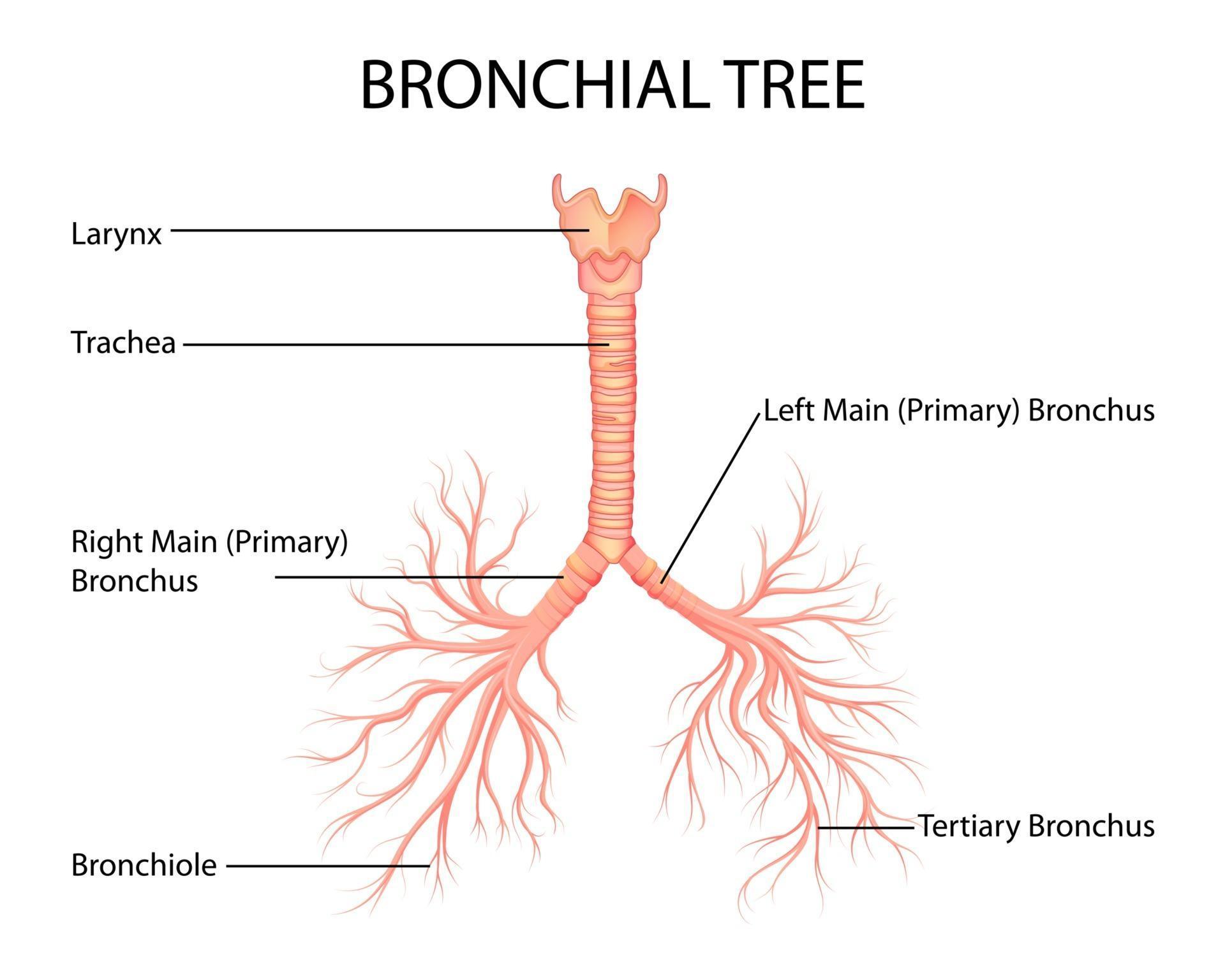
Bronchial Tree Labeled Diagram

Illustration Of Healthcare And Medical Education Drawing Chart Of Human Trachea the trachea, also called the windpipe, is part of the passageway that supplies air to the lungs. any prolonged blockage, even for a few minutes, can cause death. the trachea is about 4. The trachea, bronchi and bronchioles form the tracheobronchial tree – a system of airways that allow passage of air into the lungs, where gas exchange occurs. these airways are located in the neck and thorax. in this article we will look at the anatomical position, structure and neurovascular supply of the airways; as well as considering.

Bronchial Tree Bronchi. bronchi are plural for bronchus and represent the passageways leading into the lungs. the first bronchi branch from trachea, and they are the right and left main bronchi. these bronchi are the widest and they enter the lung. after entering the lungs, the bronchi continue to branch further into the secondary bronchi, known as lobar. Figure 19.5.1 19.5. 1: trachea (a) the tracheal tube is formed by stacked, c shaped pieces of hyaline cartilage. (b) the layer visible in this cross section of tracheal wall tissue between the hyaline cartilage and the lumen of the trachea is the mucosa, which is composed of pseudostratified ciliated columnar epithelium that contains goblet cells. Your bronchi work with your respiratory system to help you breathe. when you breathe: air passes from your mouth to your trachea. your trachea divides into your left and right bronchi. the bronchi carry air into your lungs. at the end of the bronchi, the bronchioles carry air to small sacs in your lungs called alveoli. Anatomy of the lungs & tracheobronchial tree. figure 1. mediastinal surface of the right lung showing the borders of the lung. figure 2. a right and b left lung showing impressions on the mediastinal surface. figure 3. bronchopulmonary segments of the a right and b left lung. figure 4. anterior view of the lungs.

Comments are closed.